Handy Links
SLAC News Center
SLAC Today
- Subscribe
- Archives: Feb 2006-May 20, 2011
- Archives: May 23, 2011 and later
- Submit Feedback or Story Ideas
- About SLAC Today
SLAC News
Lab News
- Interactions
- Lightsources.org
- ILC NewsLine
- Int'l Science Grid This Week
- Fermilab Today
- Berkeley Lab News
- @brookhaven TODAY
- DOE Pulse
- CERN Courier
- DESY inForm
- US / LHC
SLAC Links
- Emergency
- Safety
- Policy Repository
- Site Entry Form

- Site Maps
- M & O Review
- Computing Status & Calendar
- SLAC Colloquium
- SLACspeak
- SLACspace
- SLAC Logo
- Café Menu
- Flea Market
- Web E-mail
- Marguerite Shuttle
- Discount Commuter Passes
-
Award Reporting Form
- SPIRES
- SciDoc
- Activity Groups
- Library
Stanford
Around the Bay
Popular Microprobe on Beamline 2-3 Moves to Full-time

Metal-tipped spider fangs, ancient Roman pottery shards and arsenic-sipping ferns have one thing in common. They've all been studied by the X-ray microprobe on Beamline 2-3 at the Stanford Synchrotron Radiation Laboratory.
For the past two years, the microprobe has been available part-time for SSRL users to map specific locations of chemicals on their samples. The probe was originally designed for environmental scientists to study chemical contamination, but now users include biologists and archaeologists. Due to this growing popularity, the microprobe became a permanent fixture at SSRL in early December.
"Beamline 2-3 went from being one of the least popular beamlines to being one of the top five requested, most oversubscribed beamlines," said Sam Webb, the SSRL scientist in charge of the microprobe. "Users from many fields have seen how useful the microprobe is and are eager to use it in their experiments."
The device is one of only a handful of X-ray microprobes in the nation. The other comparable microprobe on the West Coast is installed at the Lawrence Berkeley National Laboratory's Advanced Light Source.
From environmental scientists to archaeologists, SSRL users have employed the microprobe to understand the chemical composition of their samples. French researchers placed Roman pottery in front of the beam to determine how ancient workers produced a specific rust coating called "slip." Other scientists imaged the thin metal coatings that provide strength and durability to spider fangs.
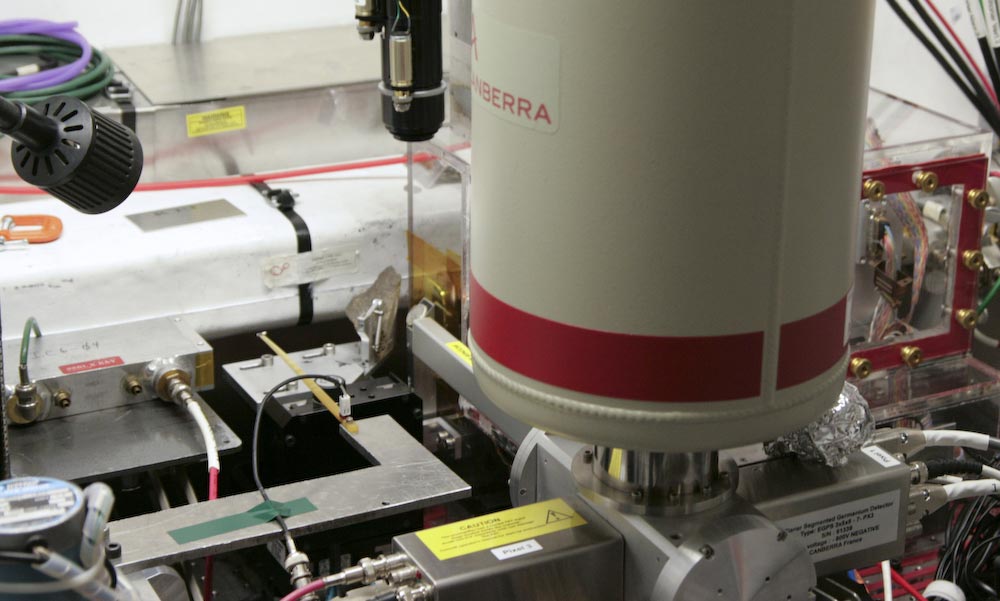
X-ray microprobes are in high demand because they can pinpoint chemical information inside extremely small areas of a sample. The SSRL device can inspect an area that is 2 microns by 2 microns. A human hair is about 100 microns thick. X-ray methodologies using larger beams can't resolve this level of detail and only collect information about the sample as a whole. They miss tiny but important structures on sample surfaces.
"These kinds of microstructures are really really super important in environmental science," said John Bargar, an SSRL scientist who helped develop the microprobe. "Even though they're very small, they can have an absolutely huge impact on chemistry that occurs in an environment." Microstructures on grains of soil in a creek or aquifer can control how toxic heavy metals leech into a water supply, Bargar said.
Five years ago, Bargar received a grant from the Department of Energy Office of Biological and Environmental Research, Environmental Remediation Science Division, to build a microprobe for studying contaminated sediments at DOE sites. He and his collaborators have since used the probe to study how soil sediments absorb uranium contaminants at different polluted sites across the country. Other environmental scientists have used the Beamline 2-3 probe to develop ways to remove uranium from ground water in Colorado and Utah.
To inspect samples at these small scales, mirrors inside the instrument narrow the incoming X-ray beam to a microscopic swatch. By moving the sample in front of the beam in two dimensions, users can expose tiny regions, one at a time.
A detector collects any light emitted from the sample. This light is created by a process called X-ray fluorescence. The incoming X-ray beam blasts electrons from the atoms inside the sample. Other electrons within the atoms fill in the holes created by those ejected, and in turn produce photons. Each element in the sample emits characteristic energies of light in this way, like a fingerprint, allowing scientists to determine which chemicals are present in the sample.
Researchers can also use Beamline 2-3 to understand the structure of the atoms inside their sample using X-ray diffraction. In these experiments, a detector placed behind the sample watches how incoming X-rays are scattered by the atoms.
In the past two years, Webb and his team have expanded the types of experiments available at the microprobe and plan to continue upgrading the equipment's capabilities. Soon users will be able to rotate their sample in the beam to create three-dimensional images.
Webb said many of the microprobe's upgrades come from users' feedback. "They tell us, 'Wouldn't it be cool if we could do this?'" Webb said. "And we say, 'Yeah that'd be cool.'"
—Michael Torrice
SLAC Today, December 8, 2008