Handy Links
SLAC News Center
SLAC Today
- Subscribe
- Archives: Feb 2006-May 20, 2011
- Archives: May 23, 2011 and later
- Submit Feedback or Story Ideas
- About SLAC Today
SLAC News
Lab News
- Interactions
- Lightsources.org
- ILC NewsLine
- Int'l Science Grid This Week
- Fermilab Today
- Berkeley Lab News
- @brookhaven TODAY
- DOE Pulse
- CERN Courier
- DESY inForm
- US / LHC
SLAC Links
- Emergency
- Safety
- Policy Repository
- Site Entry Form

- Site Maps
- M & O Review
- Computing Status & Calendar
- SLAC Colloquium
- SLACspeak
- SLACspace
- SLAC Logo
- Café Menu
- Flea Market
- Web E-mail
- Marguerite Shuttle
- Discount Commuter Passes
-
Award Reporting Form
- SPIRES
- SciDoc
- Activity Groups
- Library
Stanford
Around the Bay
Workshop Explores X-ray Effects on Biological Samples
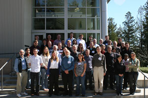
Last week more than 50 scientists from around the world came to SLAC for the Sixth International Workshop on X-ray Radiation Damage to Biological Crystalline Samples, hosted by the Stanford Synchrotron Radiation Lightsource's Structural Molecular Biology group, or SMB. The March 11—13 workshop focused on sample damage during X-ray crystallography and similar techniques used to explore the structure of proteins and other biological molecules.
Proteins serve as the tiny gears—and also transport tubes, conveyor belts and more—that keep living cells ticking. This tiny machinery is too small to see with visible light, so researchers rely on the short wavelength of X-rays to catch a glimpse of protein shapes and, from that, their likely functions. As the X-rays ripple by, the sample molecules disturb, or diffract them, creating a pattern that reveals the molecules' shape. But it takes a bright beam to create a clear diffraction pattern. The exposure can damage samples and introduce noise into already subtle data.
"Radiation damage is really a serious problem when you use very intense X-ray beams on small and weakly diffracting samples, like protein crystals," said SLAC researcher Ana Gonzalez.
Most X-ray sources require proteins to be arranged in a repeating, crystal pattern in order to get a robust view of the protein's structure. Creating these crystals can be difficult. Worse, X-rays are a very energetic form of light, capable of wreaking havoc with valuable crystallized samples. Many of last week's workshop sessions addressed means to account for and even reduce X-ray radiation's effects on samples.
"For participants actively doing research in the field of radiation damage, this is the main forum to discuss results and interchange new ideas for experiments," said Gonzalez, who co-organized the meeting with other SMB colleagues. "For beamline users, [the meeting] results in useful information about the optimal way to carry out experiments."
The launch of user science at the Linac Coherent Light Source last fall brought new elements to the workshop this year.
"A brand new topic this time was the new research opportunities offered by the developments at LCLS and similar [future] sources," Gonzalez said.
The LCLS is so bright, it is expected to image single molecules—no crystallization required. Its brilliant pulses of laser X-rays will also destroy samples, creating new challenges in data interpretation and opening a new window to the study of radiation damage.
Funding for this conference was made possible by the Department of Energy, Office of Biological and Environmental Research, the National Institutes of Health, National Institute of General Medical Sciences, Bio-X at Stanford University, Genentech, the Commissariat à l'Énergie Atomique, and by 1R13RR030726-01 from the National Center for Research Resources.
—Shawne Workman
SLAC Today, March 17, 2010
