Handy Links
SLAC News Center
SLAC Today
- Subscribe
- Archives: Feb 2006-May 20, 2011
- Archives: May 23, 2011 and later
- Submit Feedback or Story Ideas
- About SLAC Today
SLAC News
Lab News
- Interactions
- Lightsources.org
- ILC NewsLine
- Int'l Science Grid This Week
- Fermilab Today
- Berkeley Lab News
- @brookhaven TODAY
- DOE Pulse
- CERN Courier
- DESY inForm
- US / LHC
SLAC Links
- Emergency
- Safety
- Policy Repository
- Site Entry Form

- Site Maps
- M & O Review
- Computing Status & Calendar
- SLAC Colloquium
- SLACspeak
- SLACspace
- SLAC Logo
- Café Menu
- Flea Market
- Web E-mail
- Marguerite Shuttle
- Discount Commuter Passes
-
Award Reporting Form
- SPIRES
- SciDoc
- Activity Groups
- Library
Stanford
Around the Bay
Brains in the Beamline
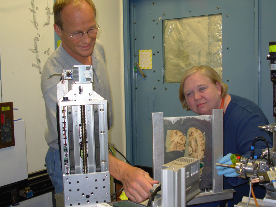 From X-rays to MRIs, advances in physics have been instrumental in improving human health. A new imaging technique developed at SLAC by Senior Staff Scientist Uwe Bergmann and his team may represent the next big advance for biological imaging. The method is currently being used to study neurodegenerative diseases, but may soon be applied to answer all kinds of medical questions.
The new method, called biological rapid scanning or X-ray fluorescence (XRF) imaging, uses the intense X-rays generated at the Stanford Synchrotron Radiation Laboratory (SSRL) to reveal the identities of trace elements in a scanned sample. The new technique is an advance over earlier microprobe X-ray analysis because it's very fast, scanning in one hour what used to take nearly 12 days to scan.
"Biological rapid scanning is complementary to other imaging techniques and should be used with other techniques," Bergmann said. XRF imaging provides lower resolution than microprobe analysis, he said, but its speed makes it practical for the first time large samples—such as the human brain—are scanned.
Rapid-scan XRF, which was first used in 2006 to scan the Archimedes Palimpsest, uses high-energy X-rays to knock electrons from the material being scanned. The material responds by emitting X-ray fluorescence. The wavelength of the emitted fluorescence reveals the identity of the elements in the scanned sample.
New software and fast read-out electronics developed at SLAC by Martin George and Alex Garachtchenko allows the X-ray beam to move continuously over the tissue sample instead of stopping after every section, saving tremendous time. The rapid scan technique, about 300 times faster than the previous method, can scan serial slices of an entire mouse brain in two hours and a slice of human brain in 10 hours. One of the first biomedical researchers to make use of this new technique was Helen Nichol from the Department of Anatomy and Cell Biology at the University of Saskatchewan. She is using the XRF imaging technique to examine human brains for traces of metal, because metal deposits are thought to play a role in neurodegenerative diseases such as Alzheimer's and Parkinson's diseases.
"XRF imaging is really opening up new vistas," Nichol said. "Each part of the brain has its own complement of metals, and with rapid-scan XRF you get to see all the elements at the same time." Metal ions are an essential component of healthy brains, Nichol said, but too much metal can be toxic and too little metal can inhibit normal cellular functions. By looking at the metal distribution in both healthy and diseased brains, Nichol hopes to gather clues to metal's role in disease. The new technique could also evaluate the safety and effectiveness of potential treatments such as metal chelation, which removes metals from the body.
XRF imaging could also lead to more effective ways to diagnose neurodegenerative diseases, said Richard McCrea, a graduate student working on the project.
"Right now most neurodegenerative diseases are clinically diagnosed through behavioral and neurological testing," McCrea said. But if brains with Parkinson's disease exhibit unusual metal deposits, "Using XRF maps as templates we could potentially improve how magnetic resonance imaging is done and make it the gold standard for Parkinson's disease diagnosis."
Studies of many other diseases can benefit from biological rapid scanning, Nichol said. Other scientists in her department are interested in using the technique to study prion diseases, epilepsy, stroke and stem cell behavior.
"It's a whole new book opening up and all we have to do is read it," Nichol said.
—Madolyn Rogers, SLAC Today, May 8, 2008
Above image: Uwe Bergmann and Helen Nichol position human brain slices for rapid scanning at SSRL. (Click on image for larger version.)