Handy Links
SLAC News Center
SLAC Today
- Subscribe
- Archives: Feb 2006-May 20, 2011
- Archives: May 23, 2011 and later
- Submit Feedback or Story Ideas
- About SLAC Today
SLAC News
Lab News
- Interactions
- Lightsources.org
- ILC NewsLine
- Int'l Science Grid This Week
- Fermilab Today
- Berkeley Lab News
- @brookhaven TODAY
- DOE Pulse
- CERN Courier
- DESY inForm
- US / LHC
SLAC Links
- Emergency
- Safety
- Policy Repository
- Site Entry Form

- Site Maps
- M & O Review
- Computing Status & Calendar
- SLAC Colloquium
- SLACspeak
- SLACspace
- SLAC Logo
- Café Menu
- Flea Market
- Web E-mail
- Marguerite Shuttle
- Discount Commuter Passes
-
Award Reporting Form
- SPIRES
- SciDoc
- Activity Groups
- Library
Stanford
Around the Bay
Big Science Gets Small
 The size of big science at the Stanford Synchrotron Radiation Laboratory (SSRL) just got a little smaller, thanks to a new X-ray microscope installed last fall at Beamline 6.
As one of only a handful of such instruments in operation around the world, the new microscope offers SSRL researchers and users a new set of eyes for peering into the three-dimensional chemistry and structure of a range of materials from meteorites to mouse bones.
The size of big science at the Stanford Synchrotron Radiation Laboratory (SSRL) just got a little smaller, thanks to a new X-ray microscope installed last fall at Beamline 6.
As one of only a handful of such instruments in operation around the world, the new microscope offers SSRL researchers and users a new set of eyes for peering into the three-dimensional chemistry and structure of a range of materials from meteorites to mouse bones.
Spearheaded by Piero Pianetta and built by X-ray equipment supplier Xradia, the otherwise commercially available microscope takes advantage of the SPEAR synchrotron's incredible brightness to quickly capture three-dimensional images of structures as small as 40 nanometers, or about the size of the smallest virus.
"It's just like a typical microscope, but instead of looking with light, which has a relatively large wavelength, we're looking with X-rays, which have a very small wavelength," said Joy Andrews, SSRL staff scientist. "That way we can look at much smaller things."
In one recent study, Andrews and colleagues investigated the effects of weightlessness on the bones of mice to gather clues about the long-term effects of spaceflight on humans. Because of the high brightness of the SPEAR synchrotron beam, scans of bone tissue can be completed in a fraction of the time needed for similar studies using other X-ray sources.
The microscope works by passing a thin beam of X-rays through the sample, then through a special lens called a "zone plate" that enlarges the image onto a special screen. This special "phosphor" screen uses the same basic technology as a traditional television screen to convert the invisible X-ray image into a visible image which is then picked up by a specially designed digital camera.
The new microscope has the added advantage of using tunable X-rays that can be changed to reveal the chemistry of a sample, giving researchers the power to scrutinize materials to a level impossible without a synchrotron X-ray source.
To obtain a final, three-dimensional image, the sample is scanned dozens of times, rotating a fraction of a degree with each shot. The images are combined to construct a movie that reveals the nano-scale structural features of the sample.
"It's identical to a CT scan, we just call it nano-tomography," said SSRL researcher Sean Brennan. "Basically we're doing CT scans of really small things."
—Brad Plummer, SLAC Today, March 4, 2008
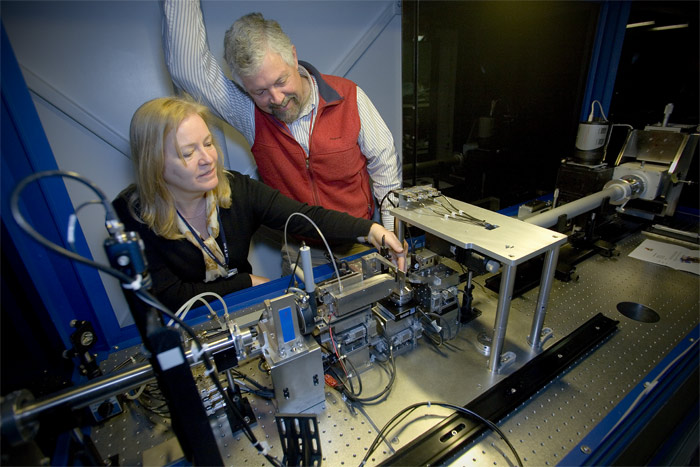
Above image: Joy Andrews (left) and Sean Brennan with the new nano-imaging X-ray microscope at SSRL Beamline 6.