Handy Links
SLAC News Center
SLAC Today
- Subscribe
- Archives: Feb 2006-May 20, 2011
- Archives: May 23, 2011 and later
- Submit Feedback or Story Ideas
- About SLAC Today
SLAC News
Lab News
- Interactions
- Lightsources.org
- ILC NewsLine
- Int'l Science Grid This Week
- Fermilab Today
- Berkeley Lab News
- @brookhaven TODAY
- DOE Pulse
- CERN Courier
- DESY inForm
- US / LHC
SLAC Links
- Emergency
- Safety
- Policy Repository
- Site Entry Form

- Site Maps
- M & O Review
- Computing Status & Calendar
- SLAC Colloquium
- SLACspeak
- SLACspace
- SLAC Logo
- Café Menu
- Flea Market
- Web E-mail
- Marguerite Shuttle
- Discount Commuter Passes
-
Award Reporting Form
- SPIRES
- SciDoc
- Activity Groups
- Library
Stanford
Around the Bay
Scanning the Microworld: SSRL's New Hard X-ray Microprobe
 Toxins like arsenic occur in soil in many parts of the world, and certain species of ferns are known for their ability to soak up these poisons. But characterizing just how they do it can be a tricky process. Now, thanks to a new microprobe at SSRL's
beamline 2-3, unlocking the secrets of how plants collect and store arsenic is getting both easier and more accurate.
Toxins like arsenic occur in soil in many parts of the world, and certain species of ferns are known for their ability to soak up these poisons. But characterizing just how they do it can be a tricky process. Now, thanks to a new microprobe at SSRL's
beamline 2-3, unlocking the secrets of how plants collect and store arsenic is getting both easier and more accurate.
Researchers have long used x-rays as a tool for studying environmental contaminants. Microprobes enable researchers to peer into the chemical structure of materials and map out the locations of compounds within a sample, such as where contaminants adhere to grains of soil. Unlike other x-ray techniques that look only at overall compositions of a sample, the microprobe allows a sample to be moved around, creating detailed picture of where compounds lie. As the x-rays scan point by point, an image emerges that shows precisely where each elements of interest resides.
The tools of the trade for x-ray researchers are often collections of instruments rather than a single, monolithic apparatus like a large microscope. The microprobe at beamline 2-3 brings together several commonly used devices, but does so in a way that gives researchers a suite of new capabilities, optimized for conducting environmental research.
A pair of focusing mirrors form the core of the new microprobe, squeezing the beam down to a two-micron spot (50 times smaller than a strand of hair and the same size as many biological cells). During a scan, a motorized sample holder precisely rasters the target back and forth across the beam, collecting points of data to create a digital image of the elements within a sample. Researchers can target specific sites on a sample with the help of a special camera that gives an x-ray's-eye view of exactly where the beam strikes.
This collection of instruments allows a unique range of approaches to data collection, but the process of analyzing the data is accomplished with custom software created for crunching the numbers as a scan proceeds. Users can point and click their way to a precise map of elements in their sample, thanks to a unique interface created by SSRL beamline scientist Sam Webb and a team of colleagues.
"Basically we're trying to streamline this process," said Webb. "A lot of work has been done to make it easier for users."
Although the new microprobe, funded by U.S. DOE BER, can in fact be tuned to study a wide variety of materials, Webb says the new suite of instruments was specifically designed to look at heavy elements like uranium. Currently, the experimental setup allows researchers to collect two kinds of data—x-ray absorption, which identifies atoms by the radiation they soak up during exposure to the beam; and x-ray fluorescence, which identifies atoms by the photons of light they emit during exposure. In the future, Webb says his team hopes to add more sophisticated detectors, such as one for conducting x-ray diffraction studies. X-ray diffraction creates images from patterns of radiation scattered onto a detector by an atom's electrons.
These techniques will help environmental and biological researchers better study the chemical makeup of materials like contaminated soil and tissue samples from plants, and in particular, to better understand the role of microstructures in the environment.
"There are several microprobes around the country," said Webb. "But there aren't that many that are dedicated to environmental contaminants, specifically uranium and similar materials. This is a very fruitful technique, and it's in high demand."
—Brad Plummer, January 12, 2007
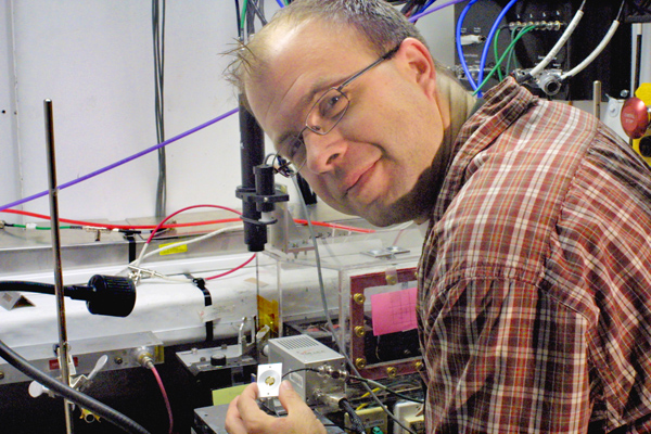
Above image: SSRL beamline scientist Sam Webb prepares to place a sample into the new hard x-ray microprobe.
Read more about arsenic imbibing ferns here.