

Monday - July 19, 2010
SLAC Today is
available online at:
http://today.slac.stanford.edu
In this issue:
Save the Date: LCLS Dedication August 16
3-D Imaging of Bone Structure Key to Understanding Bone Health
|
|
|
|
Monday - July 19, 2010 |
Save the Date: LCLS Dedication August 16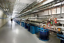 The LCLS will be dedicated on August 16, 2010.
(Photo by Brad Plummer.)
Please mark your calendars—we will be celebrating the LCLS Dedication on the afternoon of Monday, August 16th. I would like to invite each of you to take part in this celebration. Secretary of Energy Steven Chu and other guests will be attending. More details will follow as the plans unfold. This is truly a SLAC-wide accomplishment—one that would not have been possible without the support and dedication of so many people at our laboratory. I look forward to seeing you there. 3-D Imaging of Bone Structure Key to Understanding Bone Health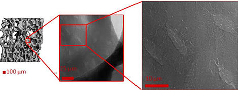 Micro-CT (left) shows trabecular structure inside bone. Transmission X-ray microscopy (center
and right) can reveal localized details. (Image courtesy
the researchers.)
The three-dimensional structure of bone is critical for maintaining strength. Skeletal diseases such as osteoporosis and environmental conditions such as weightlessness, radiation and vitamin D deficiency can affect bone structure. Understanding the 3-D structure of bone is critical to understanding how these conditions affect bone's form and function. A team that included scientists from NASA Ames Research Center, Cornell University, Xradia Inc. and the Stanford Synchrotron Radiation Lightsource has developed a technique that can be used to image bone structure in 3-D at high (30 nanometer) resolution. They used the transmission X-ray microscope on SSRL Beamline 6-2 to visualize bone from mice that had undergone a process to simulate weightlessness. Using mathematically reconstructed tomography images, they created a 3-D image of the bone's nanostructures. The scientists were also able to measure bone density on a fine scale. These techniques can be used to discern the difference between normal and diseased or distressed bone, and responses to treatment. They can also characterize nanoscale changes in bone structures due to weightlessness, and identify differences in subsequent bone regrowth upon weight bearing. This work is of particular interest to NASA as part of their research into the long-term effects of weightlessness on bone density and nanostructure. This work was published in the June issue of Microscopy and Microanalysis. To learn more about this research see the full scientific highlight. |
Events
Access (see all)
Announcements
|
|
| | ||
|
|
||
 <%
Response.AddHeader "Last-modified", getArticleDate()
'Response.AddHeader "Last-modified","Mon, 01 Sep 1997 01:03:33 GMT"
'Monday, December 06, 2010
%>
<%
Response.AddHeader "Last-modified", getArticleDate()
'Response.AddHeader "Last-modified","Mon, 01 Sep 1997 01:03:33 GMT"
'Monday, December 06, 2010
%>View online at http://today.slac.stanford.edu/. |
||
